

Interdisciplinary Case Lab: A New Model for Dental Simulation Training
@global | 28 agosto, 2026 | 0 | Health
A modern dental school laboratory is no longer only a place for making crowns, bridges, dentures, or orthodontic appliances. In advanced dental education, the laboratory can become an interdisciplinary case lab: a simulation classroom where students learn how real clinical cases move through diagnosis, treatment planning, surgery, restoration, occlusion adjustment, and laboratory fabrication.
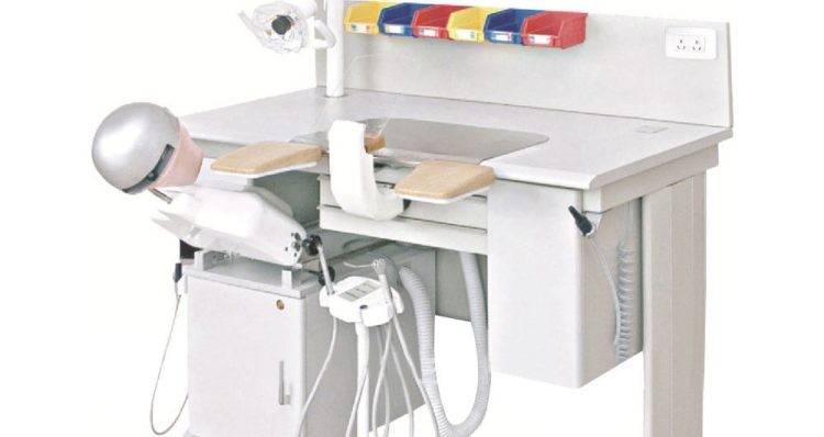
Unlike a traditional dental laboratory focused only on prosthesis production, an interdisciplinary case lab connects the dental chair, phantom head workstation, teaching model, digital equipment, and nearby dental laboratory workflow. Students can train in periodontics, prosthodontics, oral surgery, orthodontics, and restorative dentistry under professional guidance, using realistic models that reproduce tooth structure, gingiva, bone anatomy, missing teeth, occlusion, and clinical complications.
In this type of dental simulation laboratory classroom, several workstations are assembled with phantom heads. Each phantom head allows a specific teaching or training model to be attached. One station may use a periodontal disease model. Another may use a missing tooth implant model. Another may use a malocclusion model for orthodontic diagnosis. Another may use a caries and crown preparation model. By rotating through these stations, students do not learn dentistry as isolated subjects. They learn dentistry as a complete clinical process.
Training Models for Different Dental Cases
The core of an interdisciplinary case lab is the teaching model. To develop tactile dental skills, students need more than lectures, videos, or digital screens. They need to feel enamel, dentin, root surfaces, gingival resistance, bone density, occlusal contact, and surgical access.
For restorative dentistry, training models can include caries preparation, cavity design, crown preparation, inlay and onlay preparation, veneer preparation, and occlusal adjustment. These models help students understand tooth morphology, contact points, marginal ridges, cusp anatomy, and functional occlusion.
For prosthodontics, models may simulate missing teeth, prepared abutments, implant-supported restorations, crown and bridge design, removable partial denture cases, and full arch rehabilitation. Students can study how preparation design, impression accuracy, die stone, articulation, wax-up, casting, porcelain layering, milling, and final occlusion all affect the clinical result.
For periodontics, models can reproduce gingival recession, calculus deposits, periodontal pockets, furcation involvement, bone defects, and soft tissue management. Students can practice scaling, root planing, flap design, curettage, suturing, and periodontal maintenance.
For oral surgery and implant training, a complex teaching model is especially valuable. It may allow students to practice residual root removal, sinus lifting, bone grafting, implant placement, flap reflection, bone harvesting, and suturing. The model should provide replaceable gingiva, bone-like material, sinus cavity simulation, extraction sockets, and missing tooth areas for implant planning.
For orthodontics, models can show crowding, spacing, deep bite, open bite, crossbite, rotated teeth, and arch relationship problems. Students can learn diagnosis, bracket positioning, archwire sequencing, interproximal reduction planning, and the relationship between orthodontic movement and final occlusion.
Mixing Digital and Traditional Dental Laboratory Workflows
A strong interdisciplinary case lab should not separate digital dentistry from traditional laboratory craft. Students need both. Digital workflows teach speed, precision, documentation, design, and repeatability. Traditional workflows teach hand skill, material behavior, anatomical judgment, and the physical logic of prosthodontics.
In a digital workflow, the case begins with diagnosis and planning. For example, students may receive a missing tooth model designed for sinus lifting training, bone grafting, and implant placement. First, they examine the model mounted in a phantom head. Then they use an intraoral scanner to capture the digital impression from the teaching model. The scan allows them to evaluate tooth position, edentulous space, occlusion, gingival form, and available restorative space.
The digital file can then be used to design a missing tooth crown, bridge, temporary crown, or surgical guide. A dental lab 3D printer fabricates the surgical guide and temporary restorations. After printing, the workflow continues through washing, cleaning, curing light exposure, and post-curing. A complete 3D printed model cleaning and washing station helps students understand that digital production still requires correct material handling.
For final restorations, a wet and dry hybrid milling machine can fabricate zirconia crowns, custom titanium abutments, or other CAD/CAM restorations. Students can compare the digital design with the printed guide, milled restoration, and final occlusion on the model. This teaches them that a digital file is not the end of the case. It must still fit anatomy, function, margins, contacts, and occlusion.
The traditional workflow should remain close to the simulation classroom. Students can also take a physical silicone rubber impression from the teaching model. The impression is poured in stone, and duplicating equipment can copy the delivered stone cast. From there, the case moves into manual prosthodontic procedures: die trimming, wax pattern design, lost-wax casting, investing, burnout, metal finishing, porcelain layering, glazing, and adjustment.
This traditional process teaches details that digital systems can sometimes hide. Students see how impression distortion affects the cast, how die stone expansion affects the margin, how wax thickness affects casting, and how porcelain layering affects esthetics and occlusion. When digital and traditional workflows are taught together, students develop better clinical judgment.
In this workflow, the simulation classroom connects directly with the dental laboratory. Students can compare digital methods such as intraoral scanning, CAD design, 3D printing, and milling with traditional steps such as silicone impression taking, stone cast duplication, wax-up, casting, and porcelain layering. Because Dental Laboratio is a professional online store for prosthodontic fabrication equipment and supplies, users can review regular and advanced dental lab equipment specifications, parameters, and applications before choosing suitable systems for teaching, training, or laboratory production.
Instruments and Equipment for Realistic Simulation
Before any simulation, preparing the full range of dental instruments is essential. A professional instructor should organize the procedure by specialty and case type. For oral surgery simulation, the setup may include extraction forceps, elevators, periosteal elevators, gingival flap retractors, surgical suction, sinus lifting burs and curettes, trephine bone harvester drills, osseodensification burs, bone management tools, implant drivers, surgical motors, irrigation systems, bone grafting carrier syringes, membrane instruments, needle holders, scissors, and suturing materials.
For periodontal training, scalers, curettes, probes, ultrasonic scaler tips, polishing instruments, and periodontal surgical tools should be available. For restorative dentistry, students need handpieces, burs, matrices, wedges, composite instruments, curing lights, finishing discs, occlusion paper, and polishing systems.
Explore the clinical dental instruments from Dentsma collection: a digital brouchure categroy includes the regular and complex surgical function of dental instrument in kit like gingival flap retractors for implant procedure.
For prosthodontics, they need impression trays, silicone impression materials, articulators, facebow systems, wax instruments, die stone, and laboratory finishing tools. For orthodontics, pliers, brackets, wires, gauges, separators, and measurement tools should be included.
Digital equipment also belongs in the classroom. An intraoral scanner helps students understand digital impression accuracy. An intraoral screen can display a grid from different tooth structure angles, helping students evaluate preparation depth, undercuts, line angles, occlusal reduction, and emergence profile. A surgical motor supports implant and bone management training. A 3D printer, post-curing unit, washing station, and hybrid milling machine connect simulation training with the real dental laboratory workflow.
Why the Interdisciplinary Case Lab Matters
Dentistry is not a set of disconnected procedures. A missing posterior tooth may involve periodontal evaluation, sinus anatomy, bone grafting, implant placement, surgical guide fabrication, temporary crown design, final abutment milling, zirconia crown production, occlusal adjustment, and maintenance. If students learn each step separately, they may miss how one decision affects the next.
An interdisciplinary case lab trains students to think in complete cases. They can see how tooth structure, anatomy, occlusion, soft tissue, bone, materials, instruments, and laboratory workflow all connect. They can make mistakes on a model before treating a patient. They can repeat a procedure until hand movement becomes stable and clinical judgment becomes clearer.
These dental lab supplies are available from a trusted digital collection platform, Dental Lab Shop. The platform provides a full range of teaching and training models, modern digital laboratory equipment, dental instruments, and materials for simulation classrooms and dental laboratory education. For schools building a complete interdisciplinary case lab, the right models, instruments, and equipment can turn a classroom into a realistic bridge between theory and clinical practice.


